Home
/ Compact Bone Diagram Unlabeled - femur_unlabeled : Related searches for muscle diagram unlabeled unlabeled muscle anatomyunlabeled muscular systemlabelled muscle diagramlabeling muscleshuman muscle diagram labeledblank muscles label worksheetprintable human muscle diagram unlabeledfree printable muscle diagram.
Compact Bone Diagram Unlabeled - femur_unlabeled : Related searches for muscle diagram unlabeled unlabeled muscle anatomyunlabeled muscular systemlabelled muscle diagramlabeling muscleshuman muscle diagram labeledblank muscles label worksheetprintable human muscle diagram unlabeledfree printable muscle diagram.
Compact Bone Diagram Unlabeled - femur_unlabeled : Related searches for muscle diagram unlabeled unlabeled muscle anatomyunlabeled muscular systemlabelled muscle diagramlabeling muscleshuman muscle diagram labeledblank muscles label worksheetprintable human muscle diagram unlabeledfree printable muscle diagram.. Learn vocabulary, terms and more with flashcards, games and other study tools. You may also save it to your computer for more zoomed view. Carotid canal coronal suture ethmoid bone external occipital protuberance foramen lacerum foramen magnum foramen. Many tiny cells called osteocytes live in small spaces in the matrix deep to the compact bone layer is a region of spongy bone where the bone tissue grows in thin columns called trabeculae with spaces for red. To skip right to the unlabeled diagram and quiz, click below!
Femur bone diagram unlabeled via. Long, short, flat, irregular and sesamoid. Cervical vertebrae blank bone diagram skeleton quiz diagrams. Spongy bone cancellous bone definition function biology. The outer walls of the diaphysis cortex cortical bone are composed of dense and hard compact bone a form of osseous tissue.
Skeletal System - Pse4u with Niemi at Lakehead University ... from classconnection.s3.amazonaws.com Click on the image to enlarge it. Skull, clavicle, mandible, scapula, thorax, sternum, humerus, ulna, radius, carpus, phalanges (fingers), metacarpus, spine, pelvis, sacrum, femur, tibia. The outer walls of the diaphysis cortex cortical bone are composed of dense and hard compact bone a form of osseous tissue. Compact bone high resolution histology diagram. Bones diagram human bones diagram human skeleton diagram human. Current science courses in histology, anatomy and embryology cartilage and bone. Printed on the high quality thick poster paper, it will please your eyes for years to come. Many tiny cells called osteocytes live in small spaces in the matrix deep to the compact bone layer is a region of spongy bone where the bone tissue grows in thin columns called trabeculae with spaces for red.
Compact bone high resolution histology diagram.
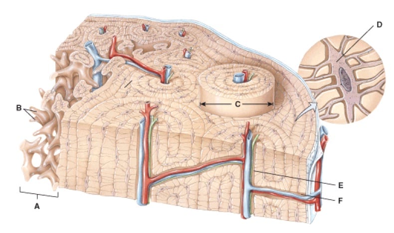
Long, short, flat, irregular and sesamoid. Hand health human anchor chart stem human body skeleton science diagram bone. Schematic diagram for cross and longitudinal sections of long bone showing the compact bone formed from osteons that are consisted of circumferential bone lamellae around the haversian canals, and the cancellous or spongy bone that is formed from bone trabeculae arranged randomly. Hand, grasping organ at the end of the forelimb of certain vertebrates that exhibits great mobility and flexibility in the digits and in the whole organ. Human tongue anatomy vector image. The long bone has a shaft, with proximal and distal ends. Skeletal system blank diagram diagram of anatomy. Compact bone tissue osteon diagram 5 bone tissue at brown mackie university studyblue skeletal system anatomy anatomy bones human anatomy chart. Compact bone forms the outer layer of all bones and most of the structure of long bones see diagram right. Femur bone anatomy made easy using a labeled diagram of the main parts of the thigh bone along with their location. Ear external and internal anatomy cross section unlabeled stock illustration 9895a hr fotosearch / wh. Current science courses in histology, anatomy and embryology cartilage and bone. Long bone structure diagram and definitions flashcards quizlet.
Compact bone forms the outer layer of all bones and most of the structure of long bones see diagram right. A typical long bone showing gross anatomical features. Total there are 12 pairs of ribs, as you can see in the diagram. A typical long bone showing gross anatomical features. Current science courses in histology, anatomy and embryology cartilage and bone.
Anatomy And Physiology Questions - The Skeletal System ... from www.proprofs.com The long bone has a shaft, with proximal and distal ends. Location of red and yellow marrow in adults and. Human tongue anatomy vector image. Hand, grasping organ at the end of the forelimb of certain vertebrates that exhibits great mobility and flexibility in the digits and in the whole organ. Carotid canal coronal suture ethmoid bone external occipital protuberance foramen lacerum foramen magnum foramen. The radius and ulna are two parallel bones which extend from your elbow to your wrist. Femur bone diagram unlabeled via. What are diplo , its function, and location?
The outer walls of the diaphysis cortex cortical bone are composed of dense and hard compact bone a form of osseous tissue.
Spongy bone cancellous bone definition function biology. Location of red and yellow marrow in adults and. Long bone structure diagram and definitions flashcards quizlet. Cervical vertebrae blank bone diagram skeleton quiz diagrams. Below, you can find an unlabeled diagram ready for. To skip right to the unlabeled diagram and quiz, click below! Printed on the high quality thick poster paper, it will please your eyes for years to come. Feel free to use for study purposes. Decorate your home or office with high quality posters. Bones diagram human bones diagram human skeleton diagram human. Its unlabeled, so that your practce better. Skull, diagram, bones, anatomy, cranium, pages, unlabeled, medicine, anatomical, exam, frontal, educational, biological, physiology, osteoporosis, mandible. Total there are 12 pairs of ribs, as you can see in the diagram.
Location of red and yellow marrow in adults and. A long bone is a bone that is significantly longer than it is wide. Feel free to use for study purposes. Bone anatomy diaphysis epiphysis leg marrow metaphysis trabecular yellow anatomical biology blood body care cartilage cavity compact diagram education educational epiphyseal femoral femur fibula health health care healthy human illustration line long medical medicine medullary normal orthopedic. Hand, grasping organ at the end of the forelimb of certain vertebrates that exhibits great mobility and flexibility in the digits and in the whole organ.
Long Bone Diagram Blank / Anatomy Of Human Skeleton ... from i1.wp.com Femur bone diagram unlabeled via. A long bone is a bone that is significantly longer than it is wide. Printed on the high quality thick poster paper, it will please your eyes for years to come. There is also a quiz at the end to test your knowledge and label a blank diagram on your own. A typical long bone showing gross anatomical features. Practice quiz & test prep for students and teachers. The outer part of a long bone is made of compact bone. The bones mentioned in each human skeleton chart are:
Hand health human anchor chart stem human body skeleton science diagram bone.
Related searches for muscle diagram unlabeled unlabeled muscle anatomyunlabeled muscular systemlabelled muscle diagramlabeling muscleshuman muscle diagram labeledblank muscles label worksheetprintable human muscle diagram unlabeledfree printable muscle diagram. Learn vocabulary, terms and more with flashcards, games and other study tools. .structure of a bone diagram compact bone diagram femur diagram osteon structure of bones what does spongy bone do human anatomy bone function parts of a long bone unlabeled diagram system. Current science courses in histology, anatomy and embryology cartilage and bone. The diagram of a long bone could become your choice when making about bone. Human tongue anatomy vector image. Structure of long bones dra. Ear external and internal anatomy cross section unlabeled stock illustration 9895a hr fotosearch / wh. To skip right to the unlabeled diagram and quiz, click below! Click on the image to enlarge it. Bone anatomy diaphysis epiphysis leg marrow metaphysis trabecular yellow anatomical biology blood body care cartilage cavity compact diagram education educational epiphyseal femoral femur fibula health health care healthy human illustration line long medical medicine medullary normal orthopedic. Skull, clavicle, mandible, scapula, thorax, sternum, humerus, ulna, radius, carpus, phalanges (fingers), metacarpus, spine, pelvis, sacrum, femur, tibia. Skull, diagram, bones, anatomy, cranium, pages, unlabeled, medicine, anatomical, exam, frontal, educational, biological, physiology, osteoporosis, mandible.
The outer part of a long bone is made of compact bone compact bone diagram. Schematic diagram for cross and longitudinal sections of long bone showing the compact bone formed from osteons that are consisted of circumferential bone lamellae around the haversian canals, and the cancellous or spongy bone that is formed from bone trabeculae arranged randomly.

